| William Crookes observed the
luminous stream, later named as cathode rays, during his experiment
using his invention known as the Crooke’s tube. While
experimenting with the cathode-ray tube, incidentally Crookes
noted that undeveloped photographic plates could be sensitized
when placed near his vacuum tube. Therefore in spite of such
peculiar observation he was not aware that such photochemical
phenomenon could be related with the radiation originated
from such rarefied tube.
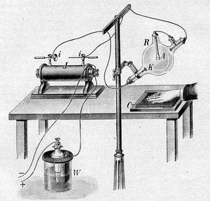 |
| Fig. 350 - An early type of X-Rays apparatus |
The history of science has many subtle situations as similar
to Crooke, another well-known physicist, Philip Lenard, did
not pay attention to investigate the fluorescent behavior
of a thin aluminum foil covered with a layer of barium platinocyanide
when hit by the cathodes rays originated from nearby Crooks
tube.
Wilhelm Conrad Roentgen was a mechanical engineer graduated
at Zurich Federal Polytechnic Institute in 1868. Even considering
he had never studied Physics he graduated with PhD through
his work about the “Study on Gases”.
Considering his great interest in Physics experiments as well
as due to his studies in exploring the behavior of the cathode
ray led him to work with others researchers as: Hertz, Hitttorf
and Crookes, and soon he was able to prove the effect of those
rays in sensitizing photographic plates.
On November 8, 1895, Roentgen repeated the experiments conducted
by Lenard using a Crookes tube provided with a type of mask.
Working in his home laboratory he noted that part of the cathode
rays generated was escaping from tube passing through the
mask. Following Lenard experiment he placed a thin aluminum
plate covered with a layer of barium platinocyanide near the
Crookes tube, observing that the invisible cathode rays had
been escaped through the mask and in this way caused a fluorescent
effect on the same.
It occurred to Roentgen that it might be advisable to open
a window in the thick glass wall of the Crookes tube in order
to allow the flow of the cathode rays. Considering the cathode
rays were invisible Roentgen noted it would be necessary to
use a kind of screen to detect their flow. Therefore, since
the cathode rays flow could be lower in the nearby wall glass
tube rather than the ones originated from the opened window
covered with thin aluminum strips, due to the intensive luminosity
in the Crookes tube probably it would be impossible to observe
the feeble fluorescence in the screen.
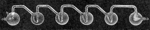 |
| Fig. 350A - In Germany, Geissler
was a forerunner in the manufacturing of vacuum tubes
provided with electrodes when connected to an electrostatic
machine or induction coil were able to generate luminous
discharged. |
In this way to avoid all the undesired environment luminosity
he covered the Crookes tube with the black cardboard hood
as well as he darkened the laboratory room.
When applying charges on the Crookes tube he observed a vivid
gleaming yellow-greenish outflow. Astonished Roentgen repeated
the experiment for several times and finally he concluded
that the fluorescence arisen was due to an unknown type of
ray, which he temporarily termed as X-Rays. Fig 350
In the reality the X-Rays are a type of electromagnetic wave
allocated in a portion of the radio frequency spectrum consisting
in a quick changing either in the electromagnetic and force
fields.
Just after the discovery of the X-Rays the future researches
were concentrated for medical application, primarily for orthopedic
diagnosis purpose only.
As usual in many discoveries the first years were conducted
in a trial and error basis considering either the precariousness
of the equipment used as well the X-Rays behavior in the human
beings was still unknown.
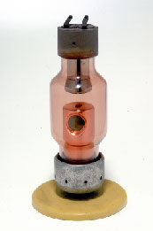 |
| Fig. 351 - A modern radiation generator,
as know as X-Rays tube. |
Therefore, due to the continuous researches after the second
half of XX century, it was possible to develop new types of
radiation generators as known as X-Rays tubes, following to
an improved calculation for proper dosing control. Fig 351
Thus to obtein good quality roentgenogram of the bone’s
tissues the X-Rays apparatus should be setup for low voltage
and current, around 70 kV.
Further to new development in the radiological techniques, the
same started to be used for diagnosis purposes in other medical
areas such as: gastroenterology, pneumatology. When diagnosing
thorax maladies, for instances, pulmonary phthisis, considering
the areas under evaluation are either in movement or even in
deeper cavities it was mandatory that the radiation generator
should operate in high tension and for short exposition time
arisen new techniques trends as the development of the fluoroscopy.
Based on such technological advancements, precision X-Rays
machines were launched in the market and when operating with
enhancing techniques physicians could detect with high accuracy
fractures, growth and many others difficulty in finding diseases.
The Roentgen’s discovery has been considered the greatest
application of the electrical phenomena in the modern medical
science and certainly it was the forerunner of the radiology
as well as the image diagnosis led to the invention in 1972
of the sophisticated computed axial tomography.
As aforementioned, the discovery of the X-rays was deeply related
with the advancements in the study of Electricity at the end
of the XX century and in this way gave birth to new fields of
researches as the ones for high accurate measuring and recording
of the feeble biological electrical currents. |
|